Abstract
STUDY DESIGN-Case-control observational study
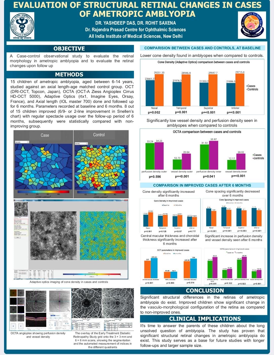
PURPOSE-To evaluate the retinal morphology in ametropic amblyopia and to evaluate the retinal changes upon follow up
METHODS–15 cases against an axial length-age matched control group. OCT, OCTA, AO, and AL done, followed up for 6 months. Parameters taken at baseline and 6 months
RESULTS-Significantly low cone density(p<0.001) and low vessel density(p<0.001) in amblyopic eyes. Upon 6 months follow up for all the case, showed significant increase in cone density(p=0.037), increase in choroidal thickness(p=0.014), Central macular thickness(p=0.001), perfusion density(p=0.022) and decrease in cone spacing(p<0.001) in the improving group
CONCLUSION-Significant structural differences in the retinas of ametropic amblyopia do exist. Improved children show significant change in the vasculo-neurological configuration of the retina as compared to non-improved ones.