Abstract
Objective: Non-contact ultra-widefield (NC-UWF) fundus imaging with integrated UWF guided swept source Optical coherence tomography (SS-OCT) was performed prospectively in preterm babies before and after intravitreal anti-VEGF (Bevacizumab) monotherapy.
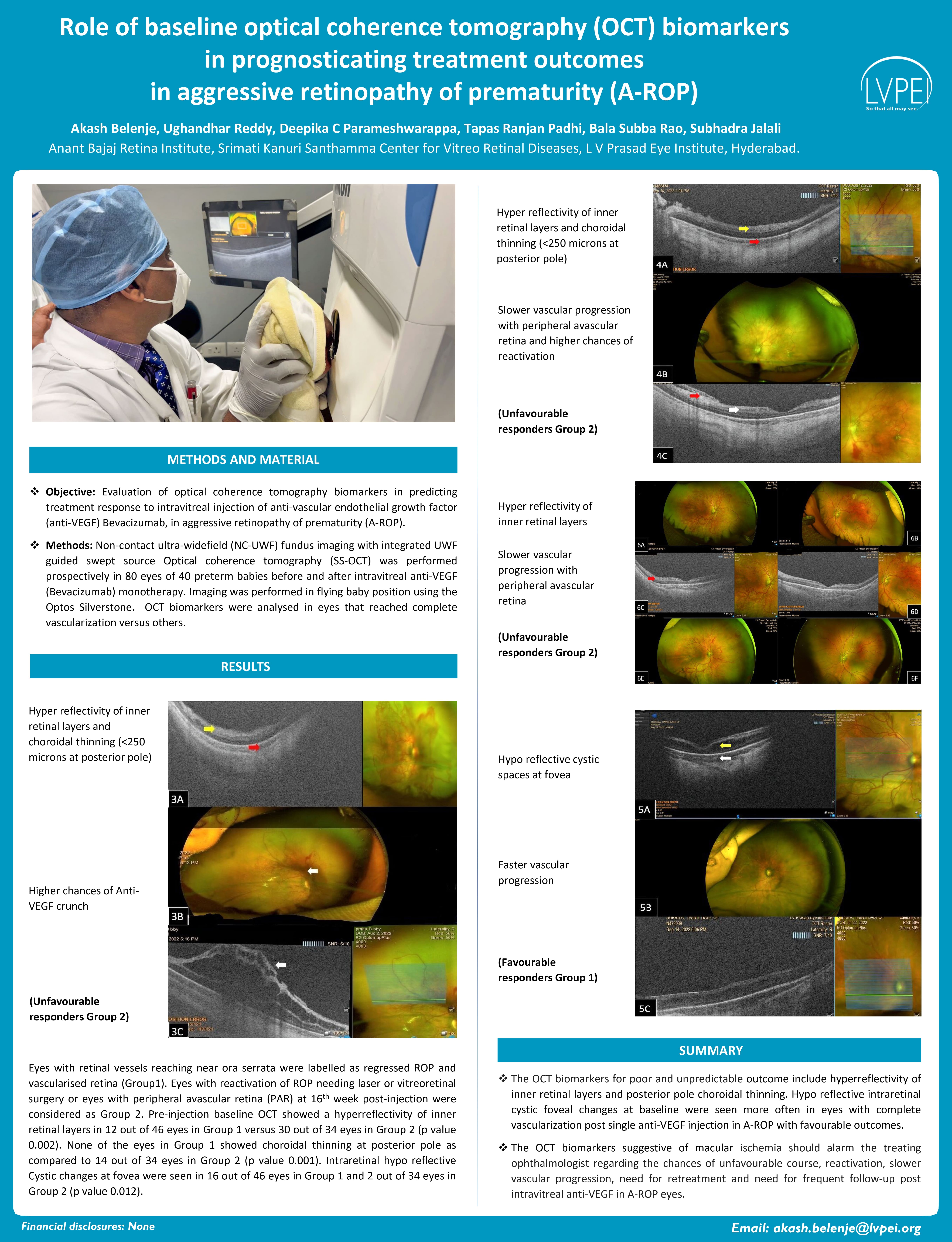
Results: Eyes with retinal vessels reaching near ora serrata were labelled as regressed ROP and vascularized retina (Group1). Eyes with reactivation of ROP needing laser or vitreoretinal surgery or eyes with peripheral avascular retina (PAR) at 16th week post-injection were considered as Group 2. Hyperreflectivity of inner retinal layers in 12 out of 46 eyes in Group 1 versus 30 out of 34 eyes in Group 2 (p value 0.002). None of the eyes in Group 1 showed choroidal thinning at posterior pole as compared to 14 out of 34 eyes in Group 2 (p value 0.001). Intraretinal hypo reflective Cystic changes at fovea were seen in 16 out of 46 eyes in Group 1 and 2 out of 34 eyes in Group 2 (p value 0.012).